Technology
Keeping up with the latest technology allows us to provide the highest standard of eyecare.
 |
Digital retinal photos
As part of the routine eye exam we will take photographs of the retina inside of your eye. This helps us to check in more detail for eye disease and we can store the images so we can refer to them next time you come. By comparing the images from previous visits we can look for subtle changes such as the early signs of glaucoma. These images have proved valuable on many occasions allowing us to detect and monitor lesions such as freckles and moles on the retina, macular degeneration and diabetes. |
|
 |
Digital corneal mapping
A corneal topogrpaher is a device that produces a 3D map of the shape and size cornea (the front surface of the eye), similar to a contour map of land or sea. Contact lenses are fitted to the cornea, so having an image of it greatly enhances our ability to accurately fit all types of contact lenses. Using this device we can better match the shape of the contact lens to the eye and now we can even create virtual contact lenses for your eyes allowing us to test a design before ordering. The topographer is also used to monitor change to the corneal shape and for the diagnosis of corneal disease such as Keratoconus which is common in New Zealand. |
|
 |
OCT (Optical Coherence Tomography)
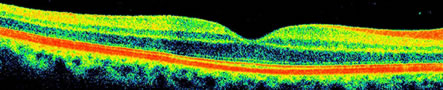 This device uses an infra-red laser to scan structures within the eye. A bit like an ultrasound, it can scan deeper into the eye tissues than we can normally see. This allows us to view to a microscopic level within or beneath the surface of a structure. It is most useful when looking for macular degeneration where fluid can sometimes accumulate beneath the surface of the retina. Also in cases of glaucoma the OCT can be used to detect and monitor the progression of damage within the optic nerve. Having an OCT image of an eye problem helps us to guide you onto the best and most timely treatment options.
|
|
|
Anterior Segment photos
With the addition of a digital SLR camera to our standard microscope, we are now able to photograph the front parts of the eye under extremely high magnification.Capturing images of lesions such as freckles and scars helps us to make comparisons in the future to tell if these lesions are changing. This camera is also useful in contact lens fitting as it allows us to record the state of a contact lens on your eye. |
|
 |
Air puff tonometer
We use a hand held "air puff" tonometer to measure the pressure inside the eye. This test is painless, requires no eye drops, and nothing has to touch your eye. In glaucoma, the eye pressure is usually elevated which is why this important test is done routinely. |
|
 |
Hoya TrueSight Dispensing Tower
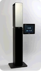 The TrueSight dispensing tower is a computerised camera and optical measuring device.
The tower takes images of you wearing your frame of choice. Using a small reference jig clipped to the frame, it can calculate extremely accurate measurements of frame/lens positions your face.
Accurate alignment of lenses in a frame is essential for high prescriptions, and progressive lens wearers. With many lenses the best vision is only achieved when looking through a specific point. This helps us to further refine the alignment of the optimum lens position with your focus.
It is frustrating when you can't see the frames you are trying on because of your high prescription. We can now use the TrueSight tower to record several images of you wearing different frames. You can then view and compare the images clearly while wearing your own glasses.
TrueSight allows you to try coloured contact lenses! After taking a photo of you, the software will digitally create simulations of you wearing different coloured contact lenses. So you can see what you will look like with different coloured eyes!
The TrueSight dispensing tower is a computerised camera and optical measuring device.
The tower takes images of you wearing your frame of choice. Using a small reference jig clipped to the frame, it can calculate extremely accurate measurements of frame/lens positions your face.
Accurate alignment of lenses in a frame is essential for high prescriptions, and progressive lens wearers. With many lenses the best vision is only achieved when looking through a specific point. This helps us to further refine the alignment of the optimum lens position with your focus.
It is frustrating when you can't see the frames you are trying on because of your high prescription. We can now use the TrueSight tower to record several images of you wearing different frames. You can then view and compare the images clearly while wearing your own glasses.
TrueSight allows you to try coloured contact lenses! After taking a photo of you, the software will digitally create simulations of you wearing different coloured contact lenses. So you can see what you will look like with different coloured eyes!
|
|
 |
Zeiss FDT Visual Field analyser
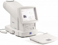 This device is to check the sensitivity of your peripheral vision. The machine is very sensitive and looks for subtle losses in peripheral vision which would be undetectable in everyday life. Peripheral vision checks are important for driving and help in the detection of eye disease such as glaucoma.
 |
|
 |
Hoya log - lens ordering system
This device links directly to our lens laboratory to enhance the ordering of spectacle lenses. While you wait it can scan your existing glasses to measure the size and curves of the lens. We can then order lenses for your existing frame without having to send your frame away to the laboratory. We can also produce 3D pictures of what your lenses will look like, showing the lens thickness which is useful to see if you have a high prescription. |
|
|
Copyright © 2004 - 2024 Highbury Optometrists Ltd. Independent eye care in Birkenhead.
